

Why Cadaver Training Matters in Medical Education
Cadaver training provides a level of anatomical realism, structural variation, and tactile experience that current digital models and virtual systems cannot fully reproduce. Unlike standardized simulations, each donor presents unique anatomy, including differences in size, vascular pathways, musculature, pathology, and tissue behavior. This exposure helps trainees build clinical judgment, develop spatial awareness, and connect theory with practice, forming a foundation for accurate diagnosis, procedural skill, and confident patient care.
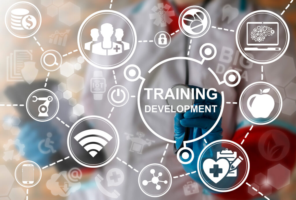
Cadaver Training in Medical and Emergency Care
Across medicine, surgery, nursing, and emergency response professions, cadaver training remains central because it immerses learners in the complexity of real human structures. Digital tools are excellent for repetition and visualization, but they cannot reliably reproduce the variability and tactile feedback encountered in real patients. Cadaver-based education strengthens clinical readiness by allowing trainees to experience tissue texture, resistance, and fragility in three-dimensional space, which supports better decision-making and sharper procedural skills. (ACS)
Why Training with Cadavers Is Still Essential Today
Real anatomy provides a level of accuracy that artificial models and animations cannot fully match. Structures vary significantly from person to person, and cadaver training exposes learners to this diversity instead of a single idealized model. Students feel tissue density, identify normal and variant anatomies, navigate around nerves and vessels, and see how disease, age, and prior surgery alter structures. This type of exposure supports more reliable clinical judgment and helps trainees translate textbook diagrams into realistic mental maps they can use with living patients. (PMC1)
Who Benefits From Cadaver-Based Education
Cadaver-based instruction supports an entire ecosystem of healthcare and emergency response professionals. Surgeons and surgical residents use cadaver labs to practice procedural sequencing, instrument handling, and approach planning before operating on patients. Physician assistants, nurses, and internal medicine residents use cadavers to rehearse procedures such as line placement, joint aspiration, and advanced physical examination techniques. Paramedics, firefighters, and other emergency medical personnel employ cadaver-based training to practice airway interventions, hemorrhage control, and ultrasound-guided procedures for rare but critical scenarios. The result is a workforce that is more technically prepared and better able to respond when seconds matter. (SAGE)
Skills Medical Trainees Develop Through Cadaver-Based Learning
Cadaver-based instruction builds a broad set of skills that directly affect patient care quality. Learners deepen their anatomical reasoning, refine surgical and procedural precision, improve their ability to interpret imaging in three dimensions, and gain confidence in responding to emergencies. Rather than practicing in isolation, trainees integrate these skills in a realistic physical context, which helps them perform more reliably when they move into clinical environments. (PMC2)
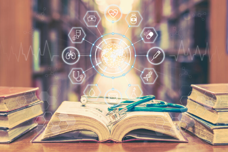
Foundational Anatomical Understanding
Dissection and cadaver-based exploration offer a depth of anatomical understanding that is difficult to achieve through images alone. Trainees see actual vascular branching patterns, organ relationships, fascia, connective tissues, and muscle compartments. They also encounter anatomical variation that would never appear in a single textbook drawing or digital model. Studies consistently show that this exposure strengthens spatial understanding and helps learners retain core anatomy for longer periods, supporting safer diagnostics and more precise procedures over the course of their careers. (RG)
Practical Procedural and Surgical Skills
Hands-on cadaver training allows learners to practice real procedures in conditions that closely resemble the operating room or trauma bay. Residents and trainees rehearse incisions, suturing, resections, catheter placements, arthroscopic access, airway procedures, and device insertion on real tissue instead of plastic. Fresh-tissue labs, in particular, offer lifelike tissue handling and realistic procedural steps. Research shows that cadaver-based curricula can improve both comfort and confidence with key procedures, such as arthrocentesis and ultrasound-guided interventions, before trainees perform them on patients. (T&F AZOrtho)
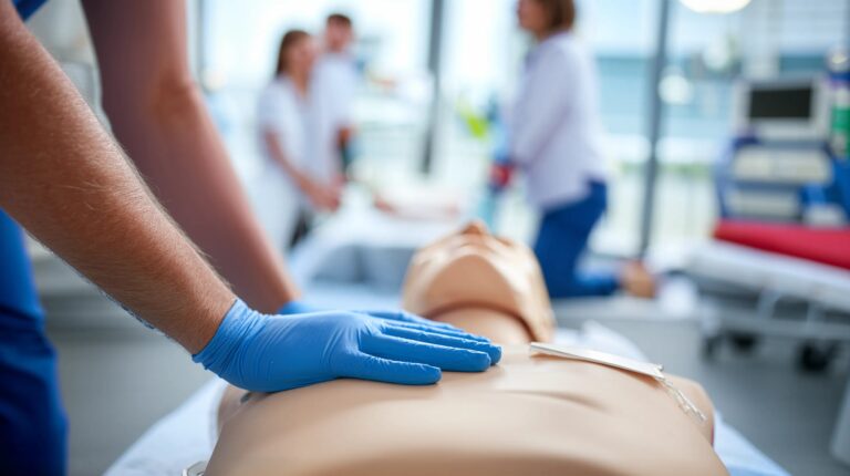
Crisis and Emergency Response Training
For emergency clinicians and responders, cadaver labs are a critical bridge between classroom training and real crises. Procedures such as trauma airway management, chest decompression, emergency cricothyrotomy, and complex hemorrhage control demand precise anatomy knowledge and composure under pressure. Cadaver-based trauma skills courses have demonstrated long-term retention of skills and confidence, which is especially important for procedures that are rarely needed but high-stakes when they are. (PubMed1)
How Cadaver Training Builds Surgical Confidence and Competence
Surgical confidence does not come from knowledge alone. It develops when clinicians have repeated opportunities to translate that knowledge into action in realistic, low-risk settings. Cadaver labs offer exactly that environment. Trainees can explore different approaches, manage unexpected anatomical findings, and refine instrument handling without the pressure of operating on a living patient. Evidence from fresh-tissue rehearsal programs shows that this preparation meaningfully increases resident confidence before live operations. (AME)
Reduced Risk and Increased Repetition
Because no live patient is at risk, cadaver labs allow learners to repeat complex procedures until they reach a safer level of competence. Trainees can slow down, analyze each step, correct errors, and receive detailed feedback from instructors. This ability to rehearse entire operations or critical segments multiple times accelerates learning and can reduce anxiety when residents perform similar procedures in the operating room for the first time.
Realistic Tissue Feedback and Technique Calibration
Current virtual tools cannot fully reproduce how real tissue stretches, tears, resists, or responds to instruments. In cadaver labs, trainees feel the difference between delicate structures and more robust tissues, learn to recognize dissection planes, and calibrate how much force to apply. This kind of tactile learning supports safer instrument use, more controlled dissection, and better protection of critical structures during surgery.
Cadaver Training vs Virtual Anatomy Tools: Why Both Matter
Virtual anatomy tools bring real advantages. They provide convenient access, allow learners to review structures from many angles, and can be used anywhere with an internet connection. However, research on anatomy education and student perceptions shows that while digital tools are valuable supplements, they do not replace the educational value of direct cadaveric dissection. For building practical skills and nuanced understanding, cadavers remain the primary reference point. (PMC3)
Strengths of Digital and Virtual Learning
High-quality 3D models, imaging platforms, and virtual dissection software make it easier to revisit complex regions repeatedly and at a learner’s own pace. These tools are especially helpful for early learners, for pre-lab preparation, and for reviewing anatomy before rotations, exams, or procedures. They also support remote learning and can reduce some logistical barriers associated with physical labs.
Limitations of Digital Tools Compared to Cadavers
Despite these advantages, digital tools are limited by their standardization. They typically present idealized anatomy and cannot show unexpected surgical findings, prior surgical changes, or subtle anatomical variation. They also lack tactile feedback, realistic tissue planes, and the sensory experience that helps learners remember how structures feel and respond. For advanced clinical and surgical training, these limitations become more noticeable.
Why a Blended Model Works but Cadavers Anchor the Learning
The most effective modern programs increasingly use a blended model. Learners use digital tools for preview, repetition, and conceptual understanding, then solidify their knowledge and skills in cadaver labs. In this model, virtual platforms enhance accessibility and review, but cadaver training anchors the learning by providing the realism required for true clinical readiness.
How Cadaver Donations Directly Support Medical Education and Patient Care
Each donated body becomes a teaching resource that shapes the skills of many future clinicians over time. Cadaver donations allow schools and training programs to offer realistic procedural practice, give residents a safe environment to learn from mistakes, and reinforce the link between anatomy and compassionate care. Recent work emphasizes that hands-on cadaveric education remains crucial for medical and surgical training even as new technologies emerge. (MDPI)
The Gift That Becomes a Lifelong Skill for Trainees
One donor may influence thousands of patient encounters. Skills learned in cadaver labs often become the techniques clinicians use for the rest of their careers. When a trainee learns how to safely perform a central line, manage a difficult airway, or execute a complex repair in a cadaver lab, each later patient who benefits from that skill is part of the donor’s legacy.
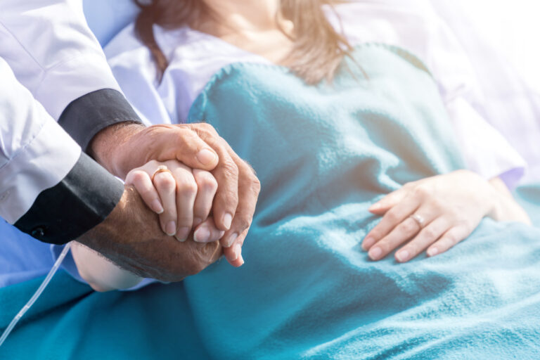
Why Donor Impact Matters for Families and Communities
For families, it can be meaningful to know that a loved one’s donation contributes to safer surgeries, better emergency response, and more confident clinicians. The impact extends beyond individual learners and touches entire communities through improved quality of care, more prepared surgical teams, and better outcomes for patients who may never know how much their providers practiced before stepping into the room.
Ethical Stewardship and Respect in Cadaver-Based Education
Respect for donors is a core principle in cadaver-based education, not an afterthought. Institutions that use cadaver labs emphasize professionalism, gratitude, and ethical conduct, and many programs formally teach students how to view donated bodies as their first patients rather than anonymous specimens.
Standards, Protocols, and Respectful Practices
Accredited programs follow strict legal, safety, and procedural standards for accepting, handling, and properly storing donated bodies. These protocols protect donor identity, maintain secure environments, and ensure that cadaver use is limited to clearly defined educational or research purposes. Regular oversight helps maintain trust in the donation process.
Honoring Donor Contributions in Medical Learning
Many schools and training programs incorporate moments of reflection or memorial events to honor donors and acknowledge the privilege of learning from them. Faculty often remind trainees that the knowledge and skills gained in the lab are possible only because of donor generosity. Survey research among anatomy faculty supports the view that cadaveric dissection promotes not just knowledge, but professional values such as empathy, respect, and responsibility toward future patients. (PMC4)
Learn more about Whole Body Donation at Research For Life’s FAQs.